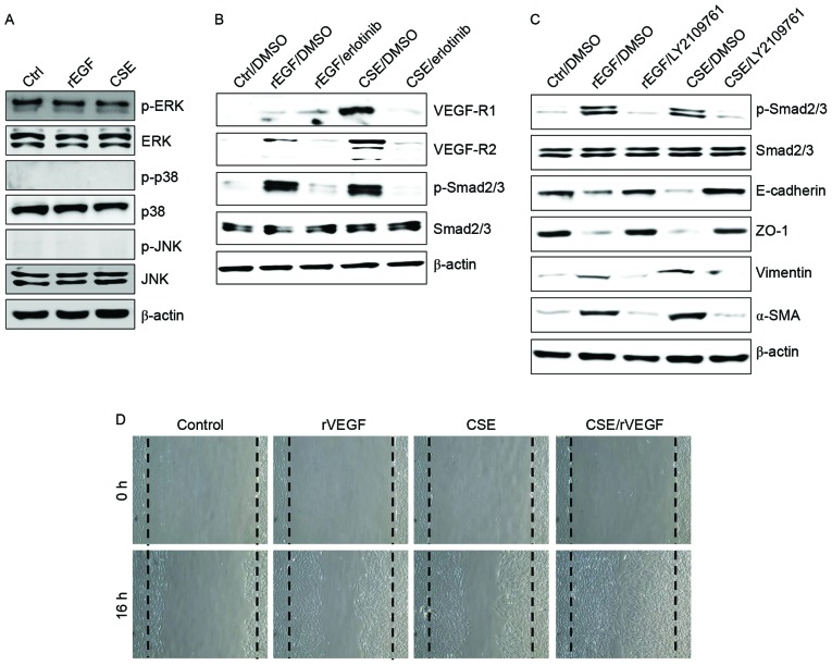
Figure 2.
CSE induces upregulation of VEGFR and activation of the transforming growth factor-β1-associated pathway. (A-C) ARPE-19 cells were exposed to 5% CSE or 100 ng/ml rEGF for 4 h, subsequently washed out, and then incubated with complete medium or 50 nM erlotinib or 100 nM LY2109761 for an additional 24 h. Total cell lysates were immunoblotted with the indicated antibodies. β-actin served as an internal control. (D) ARPE19 cells were treated with either 50 ng/ml rVEGF or 5% CSE alone, or co-treated (50 ng/ml rVEGF + 5% CSE) for 4 h, subsequently washed out, and then incubated with complete medium for an additional 24 h. Dotted lines indicate the edges of the wounds. Results are representative of three independent experiments. Ctrl, control; rEGF, recombinant epidermal growth factor; CSE, cigarette smoke extract; p, phospho; ERK, extracellular regulated kinase; JNK, c-Jun N-terminal kinase; DMSO, dimethyl sulfoxide; VEGF-R, vascular endothelial growth factor receptor; Smad, Sma- and Mad-related family; ZO-1, tight junction protein 1; α-SMA, α-smooth muscle actin; rVEGF, recombinant vascular endothelial growth factor.

