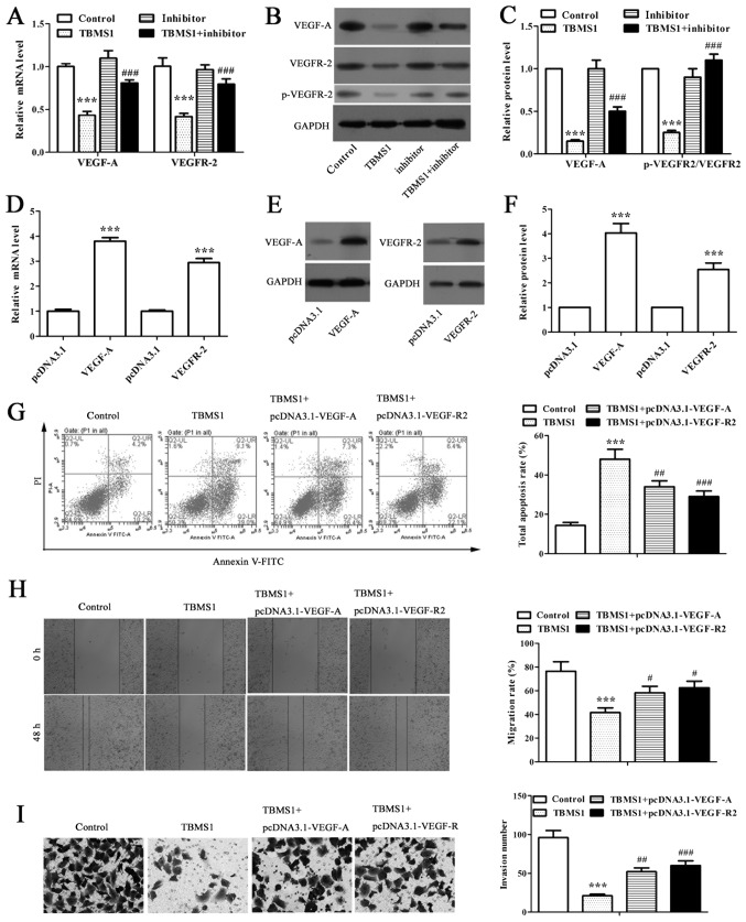
Figure 3.
TBMS1 increased miR-126-5p downregulates the expressions of VEGF-A and VEGFR-2 to enhance the apoptosis and reduce the metastasis of NCI-H1299 cells. (A) RT-qPCR analysis or (B and C) western blot analysis on VEGF-A and VEGFR-2 mRNA, protein or phosphorylation (Tyr1175) levels in TBMS1-treated NCI-H1299 cells with or without miR-126-5p inhibitor transfection. β-actin for RT-qPCR, GADPH for western blot analysis were used as internal controls. ***P<0.001 vs. control; #P<0.05, ###P<0.001 vs. TBMS1. (D) RT-qPCR analysis of VEGF-A and VEGFR-2 mRNA expression levels. β-actin was used as an internal control. ***P<0.001 vs. corresponding pcDNA3.1. (E) Representative images of western blot analysis on VEGF-A and VEGFR-2 protein expression levels. GADPH was used as an internal control. (F) Intensity comparison of western blot analysis. ***P<0.001 vs. corresponding pcDNA3.1. (G) Annexin V-FITC/PI stained flow cytometry analysis; (H) wound healing experiment (magnification, ×40); and (I) Tranwell invasion assay (magnification, ×200) were carried out in NCI-H1299 cells after 10 µmol/l TBMS1 treatment for 48 h. A series of representative images of corresponding results are shown. ***P<0.001 vs. control; #P<0.05, ##P<0.01, ###P<0.001 vs. TBMS1. All data in each group are expressed as mean ± SD from three independent experiments. TBMS1, tubeimoside-1; VEGF-A, vascular endothelial growth factor-A.

