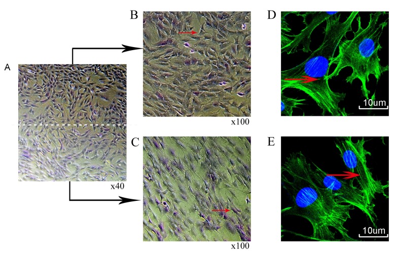
Figure 3.
Morphological changes of chondrocytes following mechanical stimulation for 3 days. (A) Toluidine blue staining (B) central and (C) peripheral regions. Phalloidin cytoskeleton staining (D) central and (E) peripheral regions; green indicates filamentous-actin; blue indicates DAPI stained nuclei.

