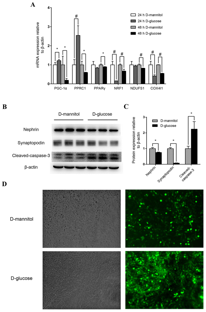
Figure 3.
Mouse podocyte and renal mesangial cell impairment by high glucose concentrations. (A) Effect of high glucose on PGC-1α, PPRC1, PPARγ, NRF1, NDUFS1 and COX4I1 mRNA expression. Mouse podocytes were cultured in 30 mM glucose medium for 24 or 48 h. Medium containing 5.5 mM glucose with 24.5 mM mannitol was applied as control. *P<0.05 and #P<0.01, as indicated. (B) Western blot analysis of nephrin, synaptopodin and cleaved-caspase-3 protein levels in podocytes following treatment with 30 mM glucose. (C) Quantification of average band intensity from four separate western blots. Data are presented as the mean ± standard deviation (n=3). *P<0.05, as indicated. (D) Levels of reactive oxygen species were detected using dichlorofluorescin diacetate and were observed with phase contrast bright-field (left panels) and epi-fluorescence (right panels) microscopy in SV40 MES 13 cells stimulated with either 30 mM D-mannitol (top) or 30 mM D-glucose (bottom) for 48 h. Magnification, ×400. PPARγ, peroxisome proliferator-activated receptor γ; PGC-1α, PPARγ coactivator-1α; PPRC1, PGC-1-related coactivator; NRF1, nuclear respiratory factor 1; NDUFS1, NADH dehydrogenase (ubiquinone) Fe-S protein 1; COX4I1, cytochrome C oxidase subunit 4I1.

