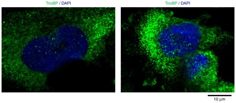
Figure 5.
Intracellular localization of TrioBP in U251-MG cell U251-MG cells were grown on glass coverslips, fixed and permeabilized with 0.2% Triton X-100. After the immuno-staining of cells with anti-TrioBP antibody, cover slips were mounted with Vectashield and visualized using a Zeiss confocal microscope. Scale bars, 10 µm.

