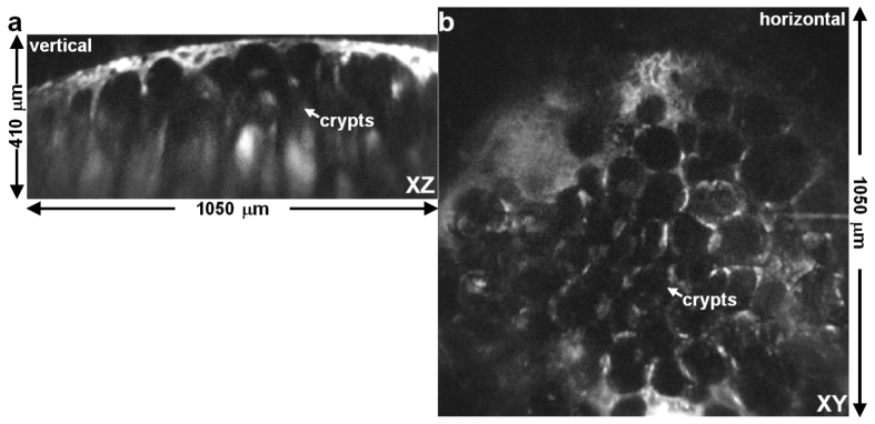
Fig. 7.
Confocal fluorescence images of human colon ex vivo. The 3D scanner was packaged in a 10 mm diameter dual axes endomicroscope to collect NIR confocal fluorescence images in either the a) vertical (XZ) plane with dimensions of 1050 × 410 μm2 or the b) horizontal (XY) plane with dimensions of 1050 × 1050 μm2. Individual crypts can be seen with either a columnar or circular shape, respectively, using IRDye800 for contrast. Image contrast was enhanced using gamma correction with a coefficient of 0.45.

