Fig. 4.
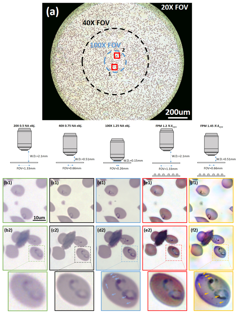
Microscope images of a malaria infected blood smear. (a) Full-sized 1.2 NAsys FPM reconstruction, which maintains the FOV and working distance of the 20X objective. The FOV of the 40X and 100X objective are marked with black and blue circles, respectively. (b1-b2) Two sub-regions from (a) (marked with red squares) captured by the 20X objective, (c1-c2) 40X 0.75 NA objective lens, and (d1-d2) 100X 1.25 NA objective lens. (e1-e2) 1.2 NAsys FPM, (f1-f2) 1.45 NAsys FPM images of cells from the same sub-regions. A malaria infected red blood cell from sub-region 2 are further zoomed in, showing particles (pointed by arrows) that are clearly resolved by 1.45 NAsys FPM and vaguely resolved by 100X oil immersion microscope.

