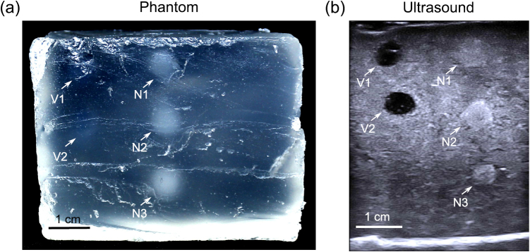
Figure 3.
Nerve and vessel phantom. (a) Photograph showing the cross-sectional view obtained after cutting through the phantom. The nerves (N1, N2, N3) are translucent, and can be visually identified from a transparent background; the vessels (V1, V2) are barely visible. (b) An ultrasound image of the phantom, in which the blood vessels present as hypoechoic and the nerves present as hyperechoic.

