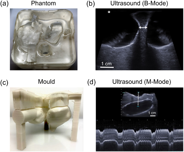
Figure 4.
Cardiac phantom. (a) Photograph (top view) without water in the heart chambers. (b) B-mode ultrasound (US) image of the septum, with the heart chambers filled with water. The septum was intact, with a minimum measured thickness of 6.6 mm (arrow). (c) The 3D printed mould used to create the phantom. (d) M-mode US image of the septum, acquired from a region indicated by the dashed line in the inset US B-mode image, which shows periodic deformation of the septum induced by manual palpation.

