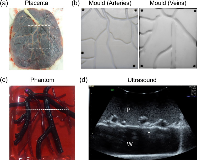
Figure 5.
Placenta phantom. (a) Photograph of the chorionic surface of a human placenta on which the phantom was based. (b) 3D printed moulds for creation of chorionic arterial and venous vasculature, derived from the human placenta (dashed region in (a); 92 mm × 99 mm). (c) Photograph of the placenta phantom. The vasculature created from the moulds shown in (b) was stretched over the rectangular placental base (94 mm × 100 mm). (d) Ultrasound image of the placenta, corresponding to the dashed white line in (c). The placenta was imaged in water, with the ultrasound probe in contact with the maternal side of the placenta, as is the case in transabdominal ultrasound imaging of the pregnant uterus. The chorionic superficial fetal vessels (arrow) were clearly apparent with hypoechoic centres and hyperechoic boundaries that were recessed from the chorionic surface of the placenta. P: placenta; W: water.

