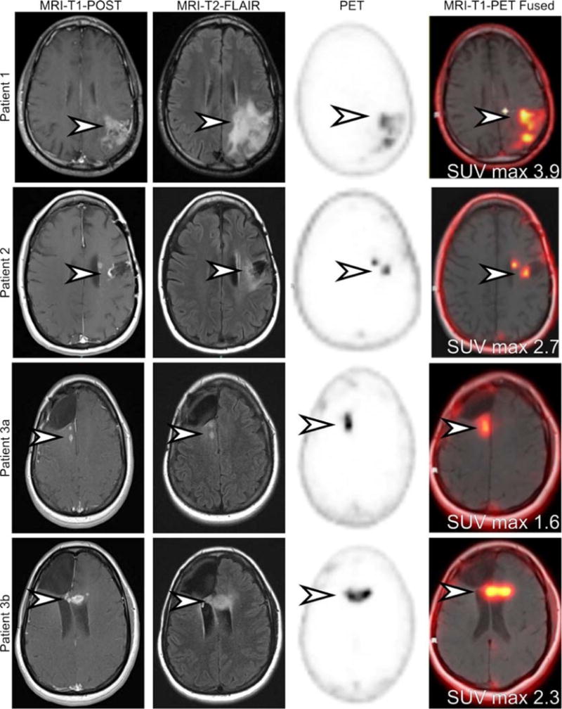
FIGURE 1.

MRI and 18F-DCFPyL PET images demonstrate areas of presumed tumor involvement and normal brain. Three patients with findings on MRI highly suggestive of glioblastoma multiforme (GBM) were prospectively recruited and imaged with [18F]DCFPyL.1,2 All patients initially had low-grade gliomas that progressed to high-grade disease and had undergone a variety of treatment regimens. Patient 1 demonstrated an area of parenchymal edema (MRI-T2-FLAIR) and MRI contrast enhancement in the frontoparietal region (arrowhead). The area demonstrated increased [18F]DCFPyL (arrowhead). Patient 2 demonstrated increased nodular frontal lobe contrast enhancement deep to a prior tumor resection cavity (arrowhead). There was associated surrounding parenchymal edema (arrowhead). The area demonstrated corresponding increased [18F]DCFPyL activity (arrowhead). Patient 3 had 2 areas of increased contrast-enhancing nodularity and parenchymal edema surrounding a prior resection cavity (arrowhead). The first lesion (3a) is located in the right periventricular white matter, whereas the second lesion (3b) is located in the genu of the left corpus callosum. Both of these lesions demonstrated increased [18F]DCFPyL activity (arrowhead). No uptake was seen in normal brain parenchyma.
