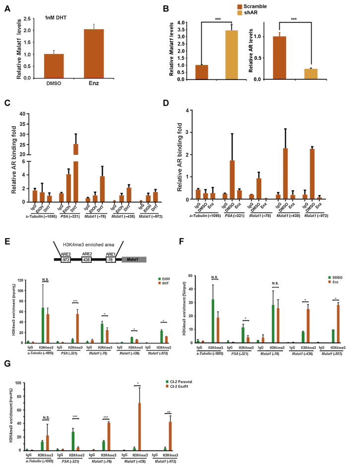
Fig. 2.
Androgen receptor (AR) regulates Malat1 expression by directly binding to its promoter. (A) C4-2 cells were cultured in media containing 1 nM dihydrotestosterone (DHT), the treatment with 10 μM enzalutamide (Enz) could result in increased expression of Malat1. (B) Suppression of AR expression in C4-2 cells through short hairpin RNA (right) increases Malat1 expression (left). (C) AR binding to regulatory elements of Malat1 promoter was enhanced in response to 10 nM DHT, and (D) was reduced in response to 10 μM Enz in C4-2 cells. Cells were treated with either 10 nM DHT or 10 μM Enz for 24 h. Chromatin immunoprecipitation assay was performed using anti-AR antibody followed by qPCR with specific primers for predicted AR binding sequence, ARE at the PSA promoter region served as positive controls and non-ARE at alpha-Tubulin promoter region was used as negative controls. (E) Top, schematic map of AR binding to Malat1 promoter and the H3K4me3 enrichment in Malat1 promoter. Bottom, DHT reduces H3K4me3 levels of Malat1 promoter after C4-2 cells were cultured with 10 nM DHT for 24 h. (F) Enz increases H3K4me3 levels of Malat1 promoter after C4-2 cells were treated with 10 μM Enz for 24 h. (G) H3K4me3 levels in Malat1 promoter are higher in C4-2 Enz-resistant cells compared to parental C4-2 cells. H3K4me3 enrichment in the promoter region of PSA or alpha-Tubulin as experimental positive or negative controls, respectively.
DMSO = dimethyl sulfoxide; IgG = immunoglobulin-G; N.S. = not significant.
* p < 0.05.
** p < 0.01.
*** p < 0.001.

