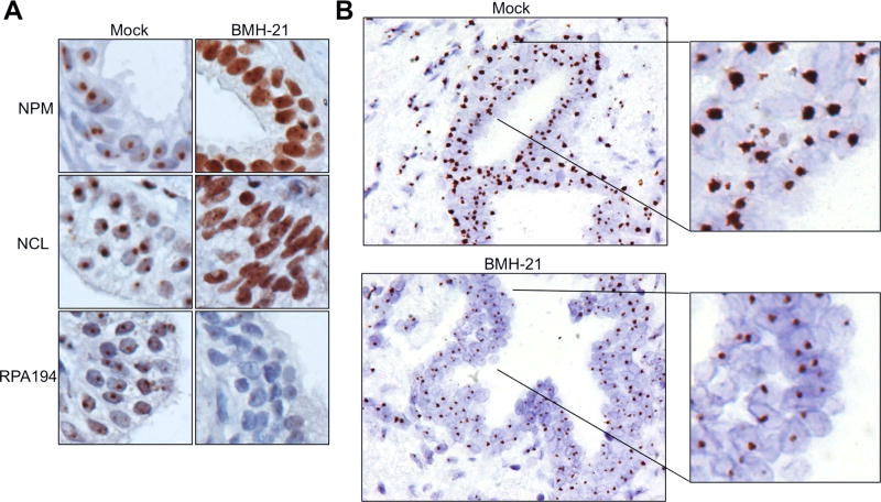
Figure 3. Suppression of 5′ETS/45S CISH signal after BMH-21 treatment of prostate tissue slices.
Tissue slices were treated with either vehicle or BMH-21 (2 µM) for 24 hours, and fixed. A, Staining of tissue slices for nucleolar markers NPM, NCL and RPA194. B, CISH for 5′ETS/45S was performed on tissues after fixing in formalin and paraffin embedding. Note marked reduction in CISH signals after BMH-21 treatment. Original magnifications, × 400; insets represent 2.8 fold magnifications.

