
Fig. 1.
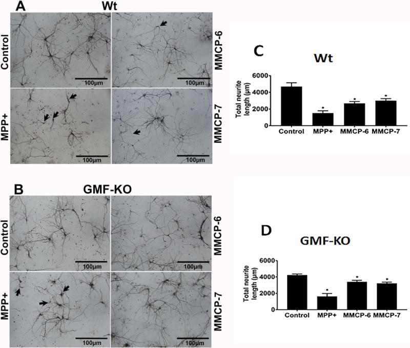
MPP+ and mast cell proteases-induce neuronal degeneration. Primary mouse neurons from Wt mice and GMF-KO mice fetal brains were cultured for 2 weeks. These cells were incubated with MPP+ (10 μM) or mast cell proteases (MMCP-6 or MMCP-7 at 100 ng/ml) for 24 h. MAP-2 ICC was performed to analyze the neuronal morphology. (A) MPP+, MMCP-6 and MMCP-7-induced significant neuronal degeneration (arrows) in the neurons obtained from Wt mice fetal brains as compared to non-treated control neuronal cells. (B) GMF-KO neurons showed less neuronal degeneration when compared to the extent of neurodegeneration observed in the neurons grown from Wt mice fetal brains. Control neuronal cells did not show any degeneration. Scale bar = 100 μm. (C, D) The total neuronal outgrowth length was measured in the whole photomicrographs using MetaMorph software to determine the neuronal degeneration. MPP+, MMCP-6 and MMCP-7 significantly reduced the total neurite length as compared with control cells as shown in the bar graphs (One-way ANOVA and Tukey-Kramer post hoc, n=3, *p<0.05). Neurodegeneration is relatively higher in Wt neurons than in GMF-KO neurons.