
Fig. 8.
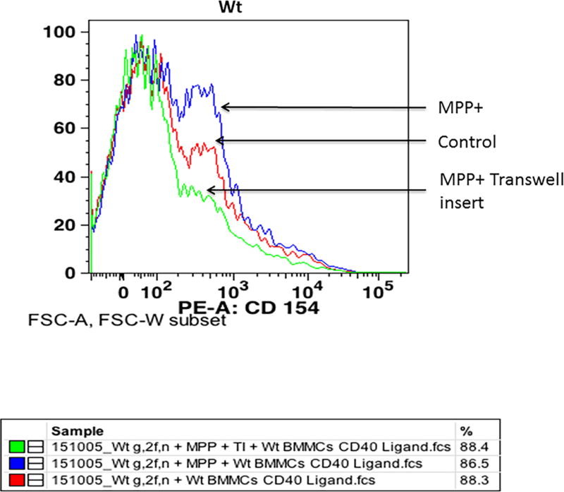
Expression of CD40L in Wt BMMCs co-cultured with glia/neurons as analyzed by flow cytometry. BMMCs and glia/neurons were co-cultured in 24 well culture plates for 72 h and subsequently incubated with MPP+ (15 μM) for 72 h at 37°C. In additional wells, transwell inserts were placed to avoid direct contact between glia/neurons and BMMCs. BMMCs were added on top of transwell inserts and incubated with MPP+. The expression of CD40L on BMMCs was analyzed by flow cytometry using anti-mCD40L/TNFSF5 antibody (n=3). MPP+ increased the expression of CD40L on BMMCs as compared to control untreated cells where there was no direct cell-to-cell contact inhibition. However, CD40L expression was reduced in the wells where transwell inserts were used to avoid direct contact of glia/neurons and BMMCs.