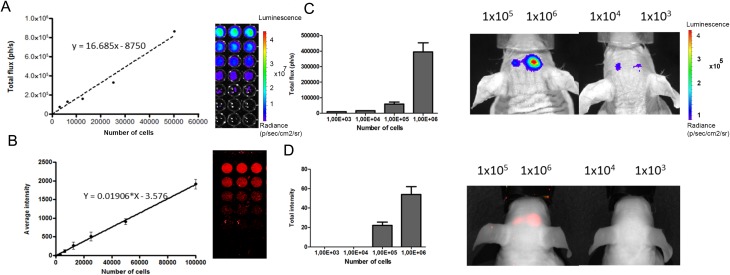
Fig. 5.
Serial dilutions of luc2-iRFP720 expressing human mesenchymal stem cells (hMSCs) measured for bioluminescence and fluorescence intensity. (A) Bioluminescence imaging (BLI) was measured for the serial dilution of of luc2-iRFP720 expressing hMSCs (0, 3.125, 6.250, 12.500, 25.000, and 50.000 cells; r 2 = 0.9921). Data are represented as mean (SD) (n = 3). (B) fluorescence imaging (FLI) was measured for the serial dilution (0, 3.125, 6.250, 12.500, 25.000, 50.000, and 100.000 cells) at 700 nm (r 2 = 0.9980) using the Odyssey scanner. Data are represented as mean (SD) (n = 6). (C) BLI of decreased amount of cells (1 × 10³, 1 × 10⁴, 1 × 10⁵, and 1 × 10⁶). Images were taken 20 min after injection of D-luciferin (25 µM/kg). (D) FLI of decreased amounts of cells (1 × 10³, 1 × 10⁴, 1 × 10⁵, and 1 × 10⁶) when injected into the mouse brain using Pearl Imager with 700 nm settings.

