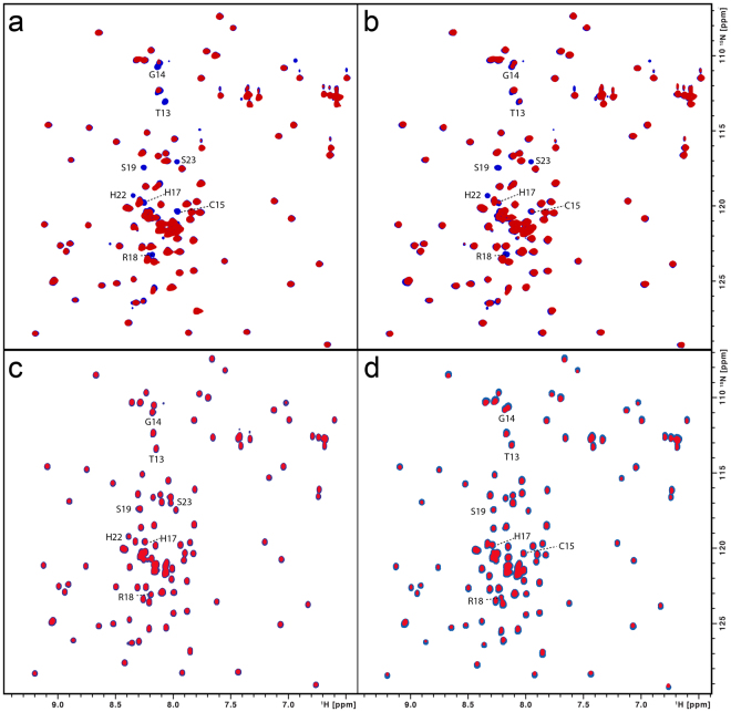
Figure 5.
(a) Superimposition of the [1H,15N]-HSQC spectra of the GB1CBS(1–40) fusion protein (100 μM) without (blue) and with hemin (50 μM; red). Interacting residues are indicated. (b) Superimposition of the [1H,15N]-HSQC spectra of the GB1CBS(1–40) fusion protein (100 μM) without (blue) and with Ga-PPIX (100 μM; red). Interacting residues are indicated. (c) Superimposition of the [1H,15N]-HSQC spectra of the C15S mutant fusion protein (100 μM) without (blue) and with hemin (50 μM; red). (d) Superimposition of the [1H,15N]-HSQC spectra of the H22L mutant fusion protein (100 μM) without (blue) and with hemin (50 μM; red).

