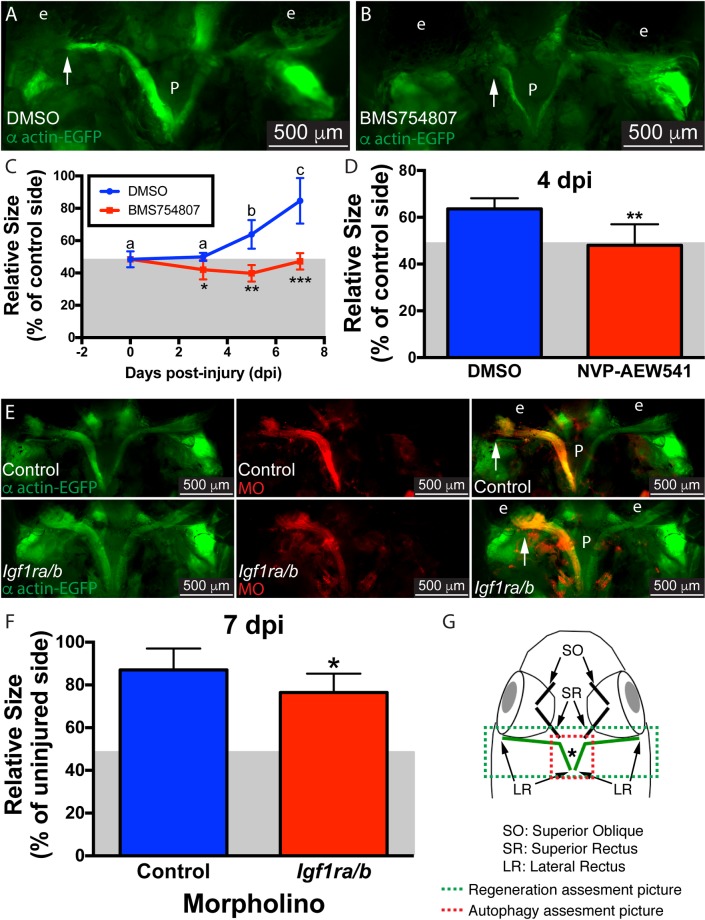
Fig 1. Inhibition of Igf1r impairs muscle regeneration.
Myectomized α-actin-EGFP fish treated with the Igf1r inhibitor BMS754807 (B) or DMSO (A) for 5 days. At selected time points (3, 5, and 7 dpi), the length of the regenerating muscle was measured as described (C), values are averages ± SD (n = 5–6). For each group (DMSO or BMS754807), differences among time points were analyzed by ANOVA. Different letters (lower case over DMSO group, there was no statistically significant difference for the BMS754807 group) indicate significant differences among time points (P < 0.05, Newman-Keuls multiple comparisons test). For each time point, differences between DMSO and BMS754807 treated fish were analyzed by Student’s t-test (*p < 0.05; **p < 0.01; ***p <0.001). To confirm our findings, α-actin-EGFP were treated with the unrelated NVP-AEW541 Igf1R inhibitor. At 4 dpi the regenerating muscle was measured as before showing similar results (D); values are averages ± SD (Student's t-test, **p < 0.01, n = 5). To knock down Igf1r, lissamine-tagged MOs (red) against both Igf1r paralogs (a and b) were microinjected into α-actin-EGFP (green) fish muscles prior myectomy. MOs were detected through the whole regenerating muscle, including the distal ends (arrowhead). Control MO (up) and Igfra/b MO (down) injected fish are shown (E). The length of the regenerating muscle was measured as described (F); values are averages ± SD (Student’s t-test, *p < 0.05, n = 10). Diagram of a craniectomized zebrafish head (G); muscles visualized by this technique are shown, and LR muscles are highlighted in green. Green and red boxes show approximately the picture used for regeneration or LysoTracker Red and GFP-Lc3 (shown in Fig 2) assessment, respectively. The white arrows mark the growing end of the regenerating muscle. P, pituitary; e, eye. Gray box in panels C, D and F represent the 50% muscle length as baseline following myectomy.

