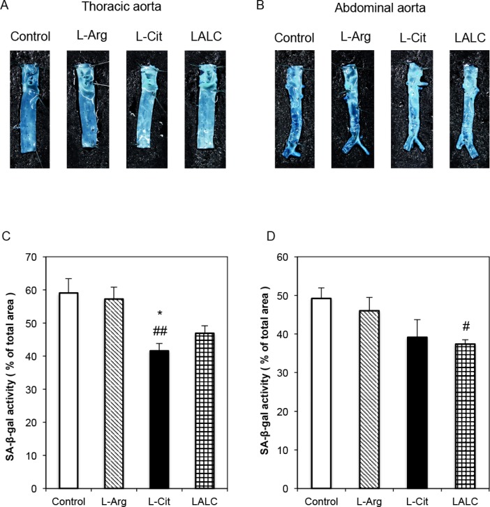
Fig 5. SA-ß-gal activity in the blood vessels of diabetic ZDFM rats.
L-Arg, L-Cit and LALC supplementation for 4 weeks reduced SA-ß-gal staining. Representative photographs of SA-ß-gal-positive staining on the intimal side of the thoracic aorta (A) and abdominal aorta (B). The relative ratio of SA-ß-gal positively stained cells on the intimal side of the thoracic aorta (C) and abdominal aorta (D). Data are expressed as the mean ± SEM. (n = 6). *P<0.05 versus Arg. ##P<0.01, #P<0.05 versus Control.

