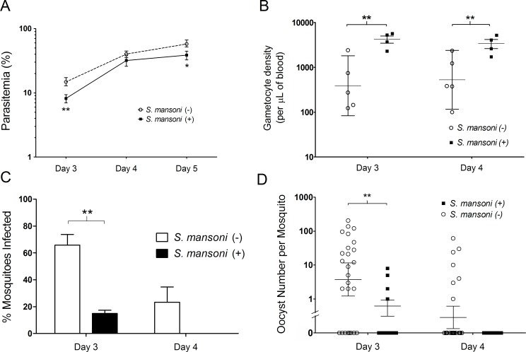
Fig 7. Gametocyte infectivity.
(A) Parasitemia. Female BALB/c mice were each inoculated with 1 x 10⁶ Plasmodium yoelii parasitized erythrocytes intravenously with (n = 4 mice) or without (n = 5) pre-existing Schistosoma mansoni infection. Parasitaemia was determined by microscopic examination on days 3, 4, and 5 post-inoculation; day 3: Student’s two-tailed t-test; **P<0.01, t = -4.906, df = 7; Day5: *P<0.05, t = -2.922, df = 5 (B) Gametocyte density. Gametocyte density was determined on days 3 and 4 post-inoculation of 1 x 10⁶ P. yoelii-parasitized erythrocytes intravenously. Day 3: Student’s two-tailed t-test; **P<0.01, t = 3.813, df = 5; Day 4: **P<0.01, t = 3.608, df = 5. Error bars show the geometric mean with 95% confidence intervals. (C) Percentage of mosquitoes with one or more oocysts present on the midgut eight days post-feeding on infected mice. A minimum of eight mosquitoes were allowed to feed on each individual mouse in the group per day **P = 0.0003, (2-way ANOVA, F = 22.23, DFn = 1, DFd = 14). Error bars mar the standard error of the mean per mouse group. (D) Oocyst numbers per mosquito. The numbers of oocysts present on mosquito midguts were determined eight days post-mosquito feeding; day 3: Student’s two-tailed t-test, **P<0.01, t = 3.077, df = 25. Error bars show the geometric mean with 95% confidence intervals. Data is representative of three independent experiments.

