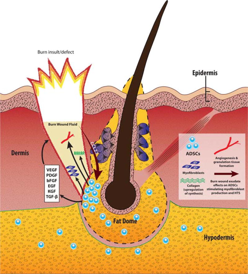
Figure 1.
The dermal cone/fat dome structure and its role in burn-related HTS pathogenesis. Burn wounds that reach the depth of the fat dome structure are more prone to develop HTS. The fat dome is thought to be a main reservoir of ADSCs. Interactions of the burn wound fluid with ADSCs shape the healing process, and in some instances direct it down the HTS pathway. Via chemokine CCL27, burn wound fluid influences residual ADSCs in the fat dome to secrete a variety of growth factors that promote collagen synthesis, angiogenesis and granulation tissue formation in the wound bed. Additionally, fat dome ADSCs are one of the putative sources of aggressive myofibroblasts that are highly characteristic of HTS. Abbreviations: HTS - hypertrophic scarring; ADSCs - adipose-derived stem cells; VEGF - vascular endothelial growth factor; PDGF - platelet-derived growth factor; bFGF - basic fibroblast growth factor; EGF - epidermal growth factor; KGF - keratinocyte growth factor; TGF-β - transforming growth factor beta; CCL27 - chemokine CCL27. [Color figure can be viewed in the online issue, which is available at wileyonlinelibrary.com.]

