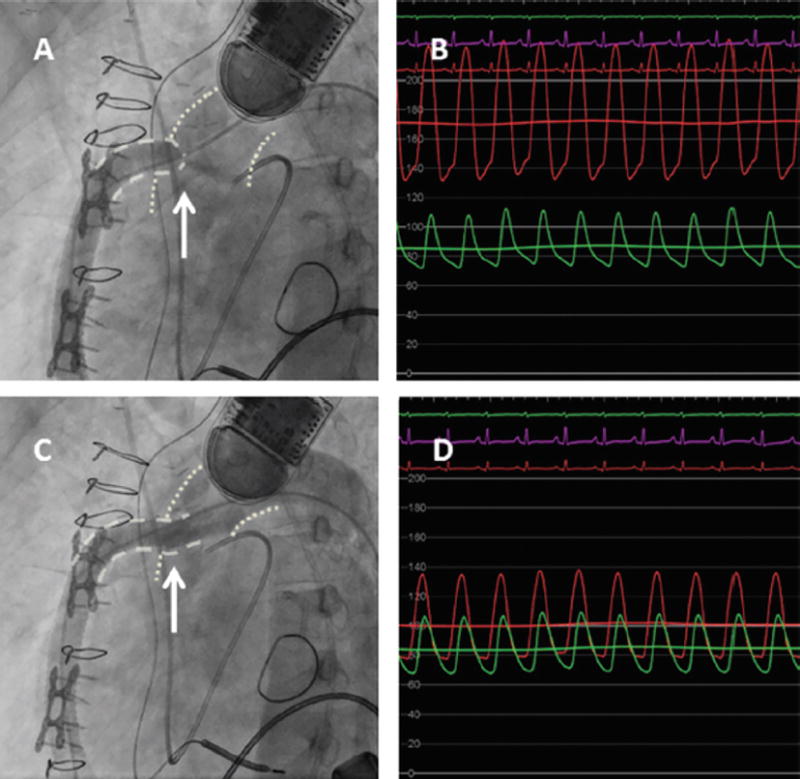
FIGURE 6.

Outflow graft stenosis in a patient with left ventricular assist device, treated with an Atrium 10 × 38 mm covered stent. (A) Significant stenosis at the aortic-outflow graft anastomosis seen on the angiogram with the arrow pointing to the tightest area of stenosis with narrow contrast jet extravasation into the aorta. (B) Hemodynamic assessment demonstrating a significant gradient across the lesion with green aortic waveform and the red waveform measured directly in the outflow graft. (C) Angiography post deployment of the Atrium 10 × 38 mm covered stent. (D) Significant improvement of the hemodynamics, from 120 mm Hg gradient to 30 mm Hg gradient. Reprinted with permission from Retzer et al.78
