
Fig. 9.
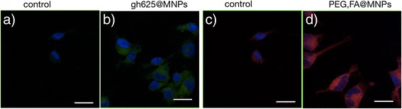
LSM fluorescence micrographs of glioblastoma cells. LSM fluorescence micrographs of glioblastoma cells. Merged blue-green images: control cells (a) and cells incubated with 15 μg/ml of NBD-labeled gH625@MNPs (b). Merged blue-red images: control cells (c) and cells incubated with 15 μg/ml rhod-labeled PEG,FA@MNPs (d). Scale bar = 20 μm