Figure 4.
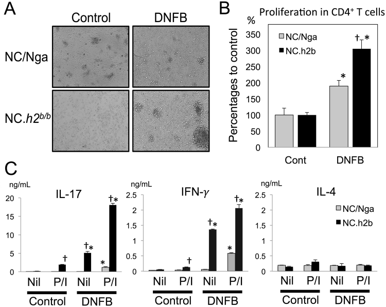
In axillary lymph nodes (LNs) with DNFB-induced dermatitis, the production of IFN-γ and IL-17 from autoreactive CD4+ T-cells in NC.h2b/b is increased compared with that in NC/Nga. The autoreactive proliferation of CD4+ T-cells purified from axillary LNs was analyzed by co-culturing with naïve CD11b+ myeloid cells from the same strain. Growth picture (A) and graph of MTT assay (B) are indicated. *DNFB vs. control, p < 0.05, ANOVA. †NC.h2b/b vs. NC/Nga, p < 0.05, ANOVA. (C) The levels of IL-17, IFN-γ and IL-4 in culture supernatant was analyzed by the ELISA method. P/I; PMA+ Ionomycin. *DNFB vs. control, p < 0.05, ANOVA. †P/I vs. Nil, p < 0.05, ANOVA. The bar graphs (n = 4) are indicated as mean ± SD.

