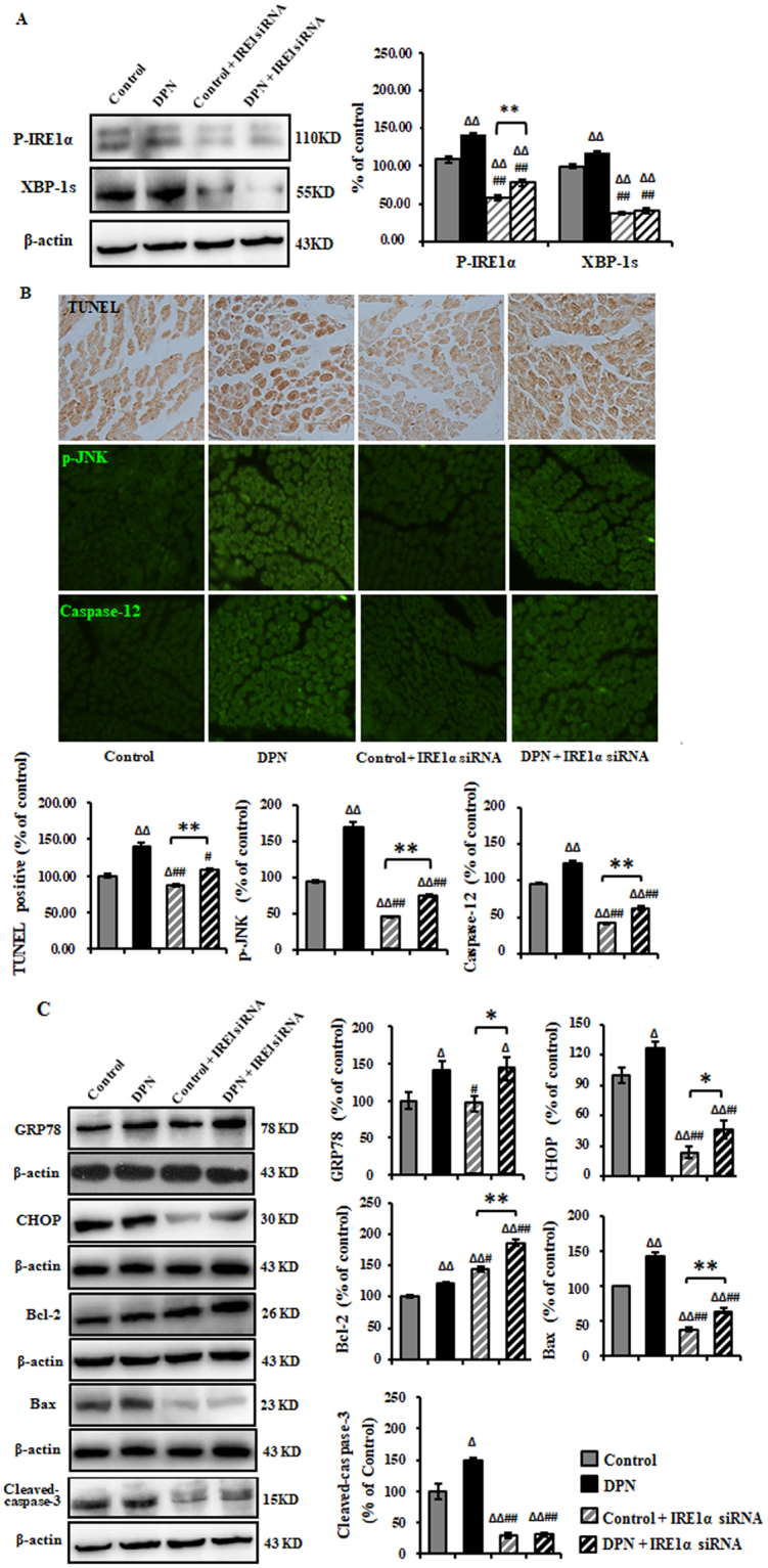
Figure 3.

Inhibition of ER stress-related apoptosis in the sciatic nerve of DPN rats by intrathecal injection of IRE1α siRNA. (A) Representative Western blots using tissue extracts from the sciatic nerve and probed with antibodies against P-IRE1α and XBP-1s. β-actin was probed as loading control. The intensity is expressed as mean ± SEM of the percentage of the respective controls and analyzed using one-way ANOVA with LSD analysis or unpaired Student’s t-test. (B) TUNEL, p-JNK, and Caspae-12 were effectively eliminated as measured by immunohistochemistry method. Data are expressed as mean ± SEM of the percentage of the respective controls and analyzed using one-way ANOVA with Tamhane’s T2 analysis or unpaired Student’s t-test. (C) Representative Western blots using tissue extracts from the sciatic nerve and probed with antibodies against GRP78, CHOP, Bcl-2, Bax and Cleaved-Caspase-3. β-actin was probed as loading control. Results of are expressed as mean ± SEM indicated as percentage of the respective controls and analyzed using one-way ANOVA with LSD analysis (GRP78, Cleaved-Caspase-3, CHOP) or Tamhane’s T2 analysis (Bcl-2, Bax) or unpaired Student’s t-test. ΔP < 0.05, ΔΔP < 0.01, compared to control; #P < 0.05, ##P < 0.01, compared to DPN; *P < 0.05, **P < 0.01 (N = 6 per group).
