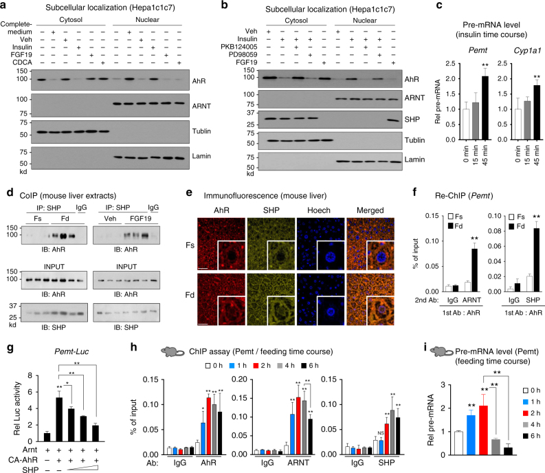
Fig. 4.
SHP inhibits AhR transactivation of Pemt in response to feeding or FGF19 treatment. a, b Hepa1c1c7 cells were grown in low-glucose and serum-free media for 12 h, and transferred for 15 min to complete medium or treated for 15 min with insulin, FGF19, or CDCA, and then cells were harvested. Cells were pre-treated with a PKB inhibitor, PKB124005, or an ERK inhibitor, PD98059, for 30 min prior to insulin treatment as indicated. Levels of AhR and SHP in the cytoplasmic and nuclear fractions were determined by IB. Consistent results were observed from two independent studies. Full size immunoblots are in Supplementary Figure 12a. c Hepa1c1c7 cells were treated with insulin for 15 min or 45 min, and pre-mRNA levels of Pemt and Cyp1a1 were measured (n = 6). d Mice were fasted for 12 h (Fs) or fasted and refed for 4 h (Fd) or fasted 4 h and treated with FGF19 for 2 h, liver nuclear extracts were prepared, and the interaction of SHP and AhR was examined by CoIP. e, f Mice were fasted overnight (Fs) or fasted and refed for 6 h (Fd). e Cellular localization of AhR and SHP in mouse liver sections was examined by IF. Scale bar: 100 µM. f Liver chromatin was immunoprecipitated with AhR antibody, then eluted, and re-precipitated with ARNT or SHP antibody to examine the occupancy of SHP at AhR-bound chromatin at the Pemt promoter (n = 5 mice). g Hepa1c1c7 cells were transfected with the indicated plasmids, and then treated with FGF19 for 2 h and luciferase activity was determined (n = 5). h, i Mice (n = 5 mice per group) were fasted for 12 h and then refed for the indicated times, and the occupancy of AhR, ARNT, and SHP at the Pemt promoter (h) or Pemt pre-mRNA levels (i) were determined by liver ChIP assay or by qRT-PCR, respectively. Means ± SD are shown, and statistical significance was measured using the c, g, i one- or f, h two-way ANOVA with the FDR post-test. *P < 0.05, **P < 0.01, NS, statistically not significant

