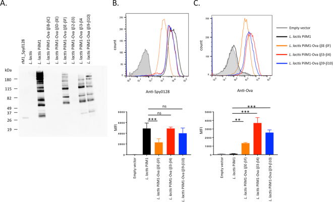
Figure 2.
Expression of the PilM1 structure on the surface of L. lactis with the model peptide Ova324–339 inserted at selected sites within the Spy0128 backbone pilus protein. (A) Western blot analysis of L. lactis cell wall extracts (CWE) with antiserum specific for M1_Spy0128 (pilus backbone protein). The high molecular band patterns are indicative of pilus assembly. For L. lactis strains that showed pilus expression after peptide insertion, flow cytometry was used to compare the expression levels of M1_Spy0128 (B) and Ova324–339 peptide (C). Error bars show the standard deviation from 3 independent experiments. **p<0.005; ***p<0.0005; one-way ANOVA followed by a Holm-Sidak test.

