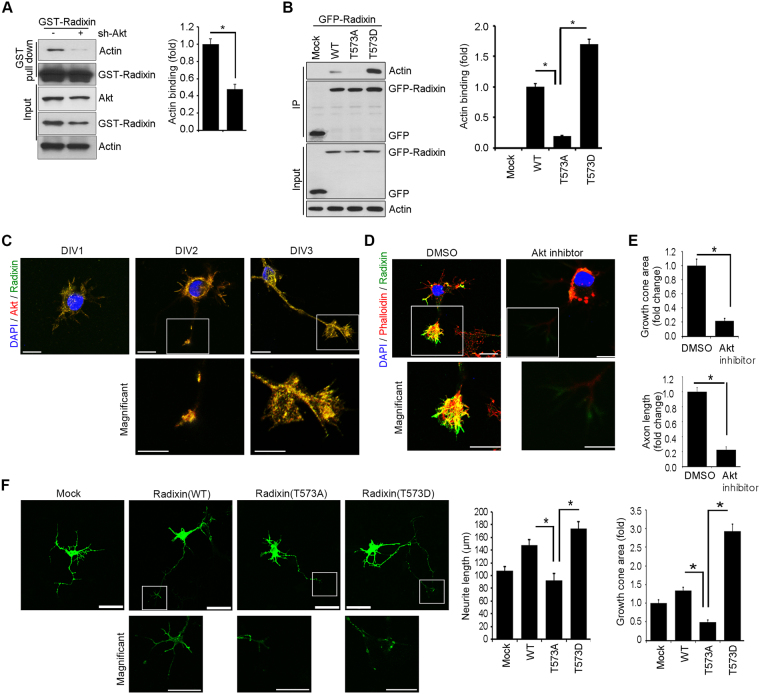
Figure 5.
Disrupting Akt-mediated radixin phosphorylation impairs radixin function in growth cone. (A) PC12 cells were depleted of Akt by sh-Akt and were transfected with GST–radixin. Lysates were then subjected to GST pull-down assay. Immunoblotting was performed with the indicated antibodies (B) Hippocampal neurons (DIV 2) were transfected with GFP−radixin−WT, radixin–T573A, or radixin–573D, and cell lysates were subjected to immunoprecipitation with anti-GFP antibody. Protein-protein interactions are determined by IB using the indicated antibodies. (A,B) Actin binding affinity was analyzed by densitometry. *p < 0.05 versus indicated. Values are mean ± SEM from three independent experiments, and the image shown is representative of at least three independent experiments. (C) The representative merged image of localization of endogenous Akt (red) and radixin (green) in the developing hippocampus neurons (DIV 1–3). Enlargement of the boxed area is shown on the bottom. Scale bar, 50 μm or 10 μm. (D) Cultured hippocampal neurons were exposed to Akt inhibitor VIII and fixed at DIV 3. The neurons stained for radixin (green) and phalloidin (red). Quantification of growth cone size and axon length measurements from three independent experiments is shown (D,E). n = 16–24 cells. Error bars, SEM; Scale bar, 10 μm or 50 μm. *p < 0.05 versus indicated. (F) Cultured neurons were transfected with GFP fusion proteins of WT radixin, radixin–T573A, radixin–T573D, or GFP vector control at DIV 1 and fixed at DIV 3. The quantification of neurite length from three independent experiments is shown (bottom). **p < 0.005 versus as indicated. Scale bar, 50 μm or 10 μm.

