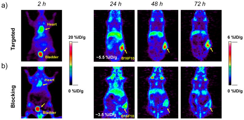
Figure 7. Serial PET imaging of 89Zr-DFO-αMSH-PEG-Cy5-C′ dots.
at 2, 24, 48 and 72 h post-injection of the MC1-R-targeting particle tracer into two cohorts of B16F10 xenografted mice: (a) targeted group and (b) NDP blocking group. For the blocking study, each mouse was co-injected with 89Zr-DFO-αMSH-PEG-Cy5-C′ dots and 200 μg of NDP. N=3 for each group.

