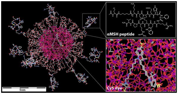
Scheme 1. Molecular rendering of αMSH-PEG-Cy5-C′ dots.
Left side shows rendering of the entire dot, with inserts on the right depicting αMSH chemical structure (top) and encapsulated Cy5 dye (bottom). Silicon, oxygen, carbon, nitrogen, sulfur and rhenium atoms are colored in purple, red, gray, blue, yellow and light green, respectively. Hydrogen atoms are not displayed in the model for better visualization.

