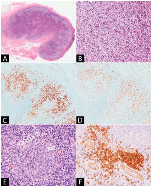
Figure 5. (A–D) Dermatopathic lymphadenitis.
(A) The lymph node paracortex is expanded by pale staining nodules. (B) The pale staining areas are composed of an admixture of dendritic cells, Langerhans cells and histiocytes, some of which contain pigment. The Langerhans cells may be numerous and are identified by immunohistochemistry with (C) CD1a and (D) langerin. (E–F) Plasmacytoid dendritic cell aggregate: (E) The plasmacytoid dendritic cells have pale cytoplasm and fine chromatin. Scattered tingible body macrophages are present. (F) CD123 stains both the plasmacytoid dendritic cells and the endothelial cells, highlighting the location of the plasmacytoid dendritic cell aggregate adjacent to the high endothelial venules.

