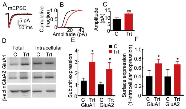
Figure 4. AMPAR-mediated neurotransmission of CA1 pyramidal neurons was also enhanced in non-epileptic animals treated with PMSG-β-HCG.
A: Averaged action potential-independent excitatory synaptic current (mEPSCs) recorded from CA1 pyramidal neuron from a control (vehicle-treated, black) and a PMSG-β-HCG-treated animal (red). B: Cumulative amplitude distribution plot of mEPSCs recorded from the representative neurons shown in A. C: Mean of the median mEPSC amplitude in the treated animals (n= 10 cells/6 hormone-treated animals and 6 cells/5 vehicle-treated animals, ** p<0.001, Student’s t-test). D: Western blots illustrating the expression of GluA1 and GluA2 subunits in the total proteins and intracellular proteins isolated from hippocampi of PMSG-β-HCG-treated animals. E: Ratio of OD AMPAR subunit expression to that of the β-actin expression, n= 9, * p<0.05, Student’s t-test). F: The surface expression of GluA1 and GluA2 subunits in the treated animals compared to that in the controls (n= 9, * p<0.05, Student’s t-test). These animals were not matched for estrous cycle.

