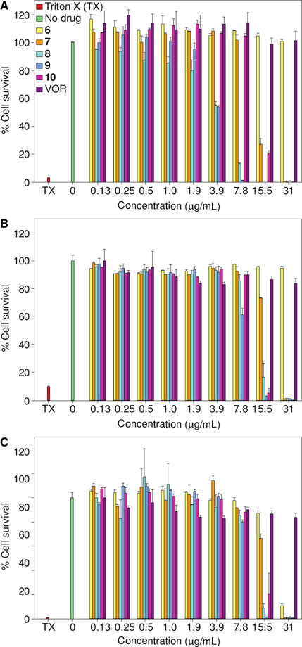
Fig. 5.

Representative cytotoxicity assays of compounds 6-10 against three mammalian cell lines: A. HEK-293, B. BEAS-2B, and C. A549. Cells were treated with various concentrations of compounds 6 (yellow), 7 (orange), 8 (turquoise), 9 (blue), 10 (pink), and VOR (purple). The positive control consisted of cells treated with Triton X-100® (TX, 20% v/v, pink). The negative control consisted of cells treated with DMSO (no drug, green). The experiments were performed in duplicate.
