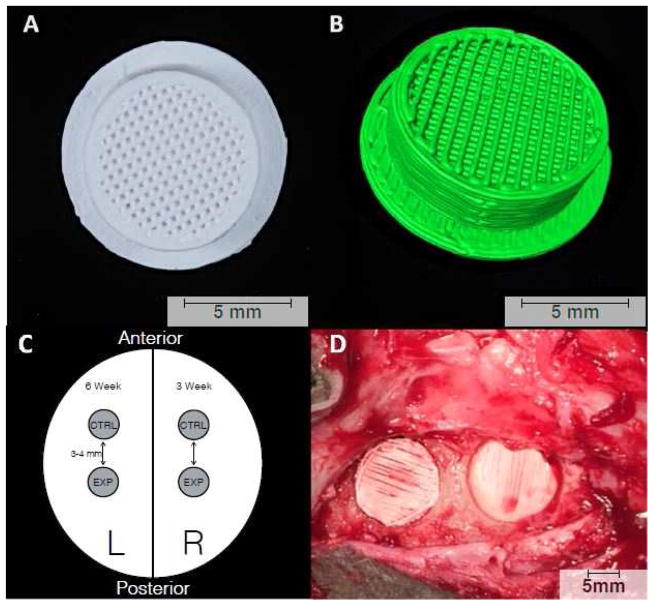
Figure 1.
3D-printed bioactive ceramic scaffold composed of β-tricalcium phosphate. (A) Inferior surface of the scaffold showcasing the porous core with central lattice. The scaffolds were placed in the trephine-induced calvarial defects such that this lattice-work faced the dura. (B) 3D reconstruction of the scaffold created using Amira 6.1 software (Visage Imaging GmbH, Berlin, Germany). (C) Schematic representation of experimental design showing location of placement for each study group and time point. (D) Intra-operative photograph showing scaffold placement in anterior and posterior calvarial defects.

