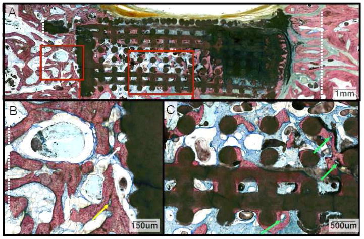
Figure 5.
(A) Representative histologic image from animals in the control group at the 6-week time point. (B) Magnification of abundant bone growth between the defect margin and wall of the scaffold (White Arrow) with a primary osteon highlighted (Yellow Arrow). (C) Along the inner lattice of the scaffold, woven bone is abundant and there is evidence of early remodeling with formation of osteons and angiogenesis (Green Arrows).

