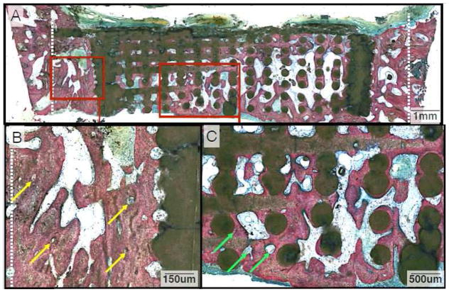
Figure 6.
(A) Representative histologic image from animals in the DIPY group at the 6-week time point. (B) Close-up depicting significant bony infill within the space between the defect margin and wall of the scaffold, with several osteons highlighted (Yellow Arrows) that provide evidence of lamellar reorganization. (C) Magnification of the extensive bone formation observed along the scaffold’s inner lattice. Angiogenesis and osteon development is evident (Green Arrows) and more prevalent in the DIPY group compared to controls at this later time point.

