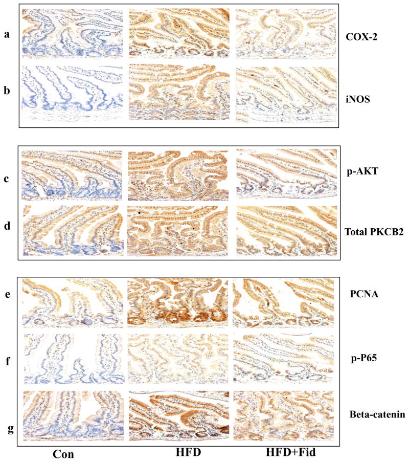
Figure 3. Inhibition of AR prevents the expression of inflammatory and pre-neoplastic markers in the small intestines of HFD-treated ApcMin/+ mice.
Histological sections of small intestines from HFD- and HFD+Fidarestat- treated mice were stained with antibodies against COX-2 (a), iNOS (b), phospho-AKT (c), PKCβ2 (d), PCNA (e), phospho- NF-κB (f) and β-catenin (g). Immunoreactivity of the antibody was assessed by dark brown staining in the intestinal cells, whereas the non-reactive areas displayed only the background color. Photomicrographs of the stained sections were acquired using an EPI-800 microscope (bright-field) connected to a Nikon camera (X400 magnification). A representative image from each group is shown. Con, control; HFD, high fat diet; HFD + Fid, high fat diet + fidarestat.

