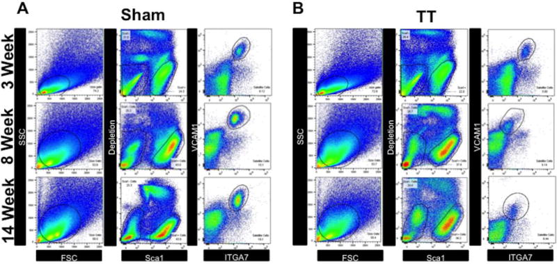
Fig. 2. Fluorescence-activated cell sorting of MuSCs from RC muscle.
(A–B) Whole RC muscle was digested into single cell suspensions and stained with anti-CD31, anti-CD45, anti-Sca1, anti-ITGA7, and anti-VCAM1 antibodies. Stained cell suspensions were then then sorted by the following schema: SSC/FSC granularity/size gating (left panels), removal of doublets (no shown), followed by depletion of CD31, CD45, Sca1 cells, and sytox positive cells (middle panels), and finally positive selection and sorting of ITGA7/VCAM1 double positive MuSCs (right panels).
(A) Representative FACS sorting gates from sham muscle at 3 weeks, 8 weeks, and 14 weeks.
(B) Representative FACS sorting gates from TT injured muscle at 3 weeks, 8 weeks, and 14 weeks.

