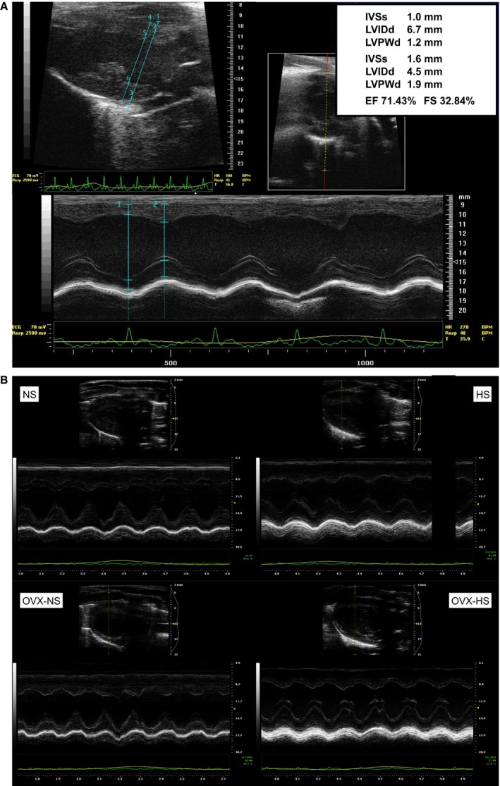
Figure 3.

(A) Echocardiographic image (parasternal long and short axis B‐mode with corresponding M‐mode image) obtained at baseline (age 12 weeks). (B) Echocardiographic images of parasternal long axis B‐mode with corresponding M‐mode obtained from each of the four groups in the study at endpoint age 28 weeks. Normal salt NS. High salt HS. Ovariectomy and normal salt OVX NS. Ovariectomy and high salt HS OVX. Normal salt NS, high salt (HS), ovariectomy with normal salt (OVX NS) and ovariectomy with HS (OVX HS).
