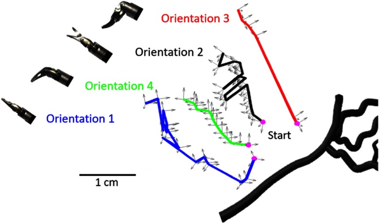
Fig. 4.
Trajectories of the scissor tool wrist with axes representing the wrist position and the da Vinci® arm orientation. These trajectories are shown relative to the vessel branch that was imaged. The ultrasound probe was located at the bottom of this image. A video showing the sweeping motion and direction relative to the phantom is included as a separate file (see Fig. 5 for more details).

