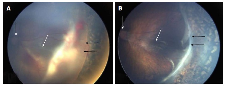
Figure 2.

Anatomical success was achieved in all ten eyes at 4-mo follow-up. A: Preoperative picture of left eyes showing stage 4A ROP with partial retinal detachment (black arrows), and the optic disc is shown by the thin white arrow; B: Postoperative picture of the same eye showing settled retinal detachment with residual fibrous tissue (black arrows). The optic disc and fovea are shown by the white arrows, respectively.
