
Figure 2.
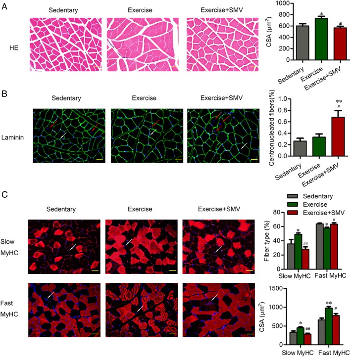
Simvastatin induced muscle damage and fibre type shift in ApoE−/− mice. ApoE−/− mice were assigned to three groups: sedentary, exercise, and exercise + simvastatin (SMV). (A) The morphologic changes of gastrocnemius were presented by haematoxylin and eosin (HE) staining. Histogram shows the cross‐sectional areas (CSA) of muscle fibres. (B) Gastrocnemius sections were stained with antibody of laminin and 40, 6‐diamidino‐2‐phenylindole (DAPI). The red arrows point to the centronucleated fibres. The white arrows point to the examples of DAPI‐labelled nuclei. Histogram shows the percentage of centronucleated fibres with respect to the total number of fibres. (C) Gastrocnemius sections were stained with antibody of slow or fast myosin heavy chain [slow myosin heavy chain (MyHC) or fast MyHC]. DAPI was used to detect nuclei. The white arrows point to the examples of DAPI‐labelled nuclei. Histograms show the percentage of fibre types with respect to the total number of fibres and the CSA of each type fibres. N = 6. * P < 0.05 and ** P < 0.01 vs. sedentary; # P < 0.05 and ## P < 0.01 vs. exercise. Scale bar: 20 μm.