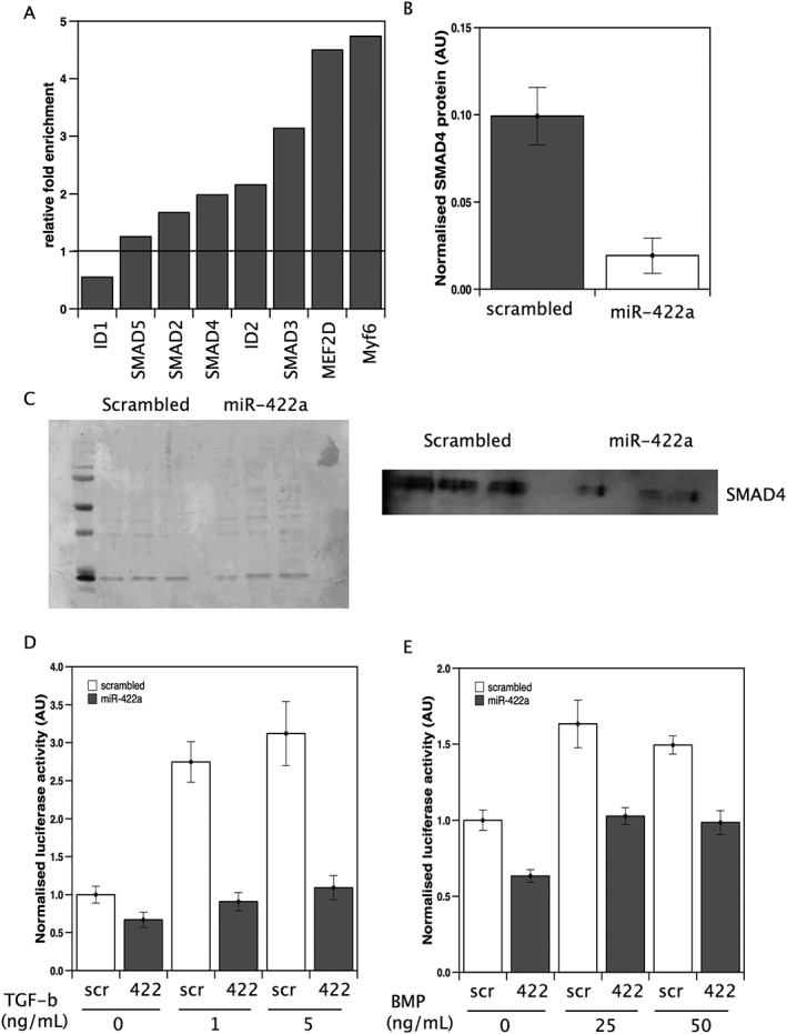
Figure 1.

miR‐422a targets SMAD4 expression in muscle cells. (A) mRNAs for predicted miR‐422a targets were quantified in RNA co‐immunoprecipitated with anti‐Ago2 from cells transfected with miR‐422a or scrambled control as described in the Methods. Fold enrichment of each target was determined by normalizing the signals to a control gene within the sample and this value for miR‐422a‐transfected cells to scrambled‐transfected cells. The data represent average fold enrichment from two independent experiments. (B and C) LHCN‐M2 cells were transfected with miR‐422a mimic or scrambled control. Forty‐eight hours later, the cells were lysed and protein extracted. Western blots were quantified, and data normalised to total protein (Ponceau S stain, C; left‐hand panel). miR‐422a caused a marked reduction in SMAD4 expression. Data are taken from three independent transfections. LHCN‐M2 cells were transfected with miR‐422a mimic or scrambled control followed by the appropriate reporter constructs as described in Methods. Cells were treated with TGF‐β (D) or BMP (E) at the stated concentrations, and luciferase activity was measured 2 h later. TGF‐β increased luciferase at both 1 and 5 ng/mL. miR‐422a inhibited the increase in luciferase activity at both doses (P = <0.001 in both cases). BMP increased luciferase at both 25 and 50 ng/mL. miR‐422a suppressed basal and ligand‐stimulated luciferase activity. Data presented are from three independent experiments performed in hextuplicate.
