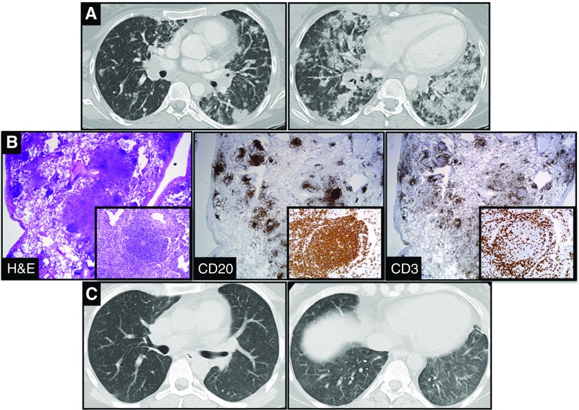
Figure 1.
(A) Coronal chest computed tomography images that demonstrated nodular infiltrates more prominent toward the lung bases in a 28-year-old woman with a 3-month history of dyspnea and a nonproductive cough. (B) Histological sections from video-assisted thoracic surgery biopsy demonstrated nodular lymphoid hyperplasia with follicles centered on small airways and areas of organizing pneumonia. Immunostains demonstrated follicles composed of CD3+ T cells surrounding a center grouping of CD20+ B cells (×20 magnification). Insets represent serial immunostains of a lymphoid follicle in the lower magnification section (×200 magnification). (C) One month after administration of azathioprine and rituximab, findings on coronal computed tomography images were dramatically improved. H&E = hematoxylin and eosin.

