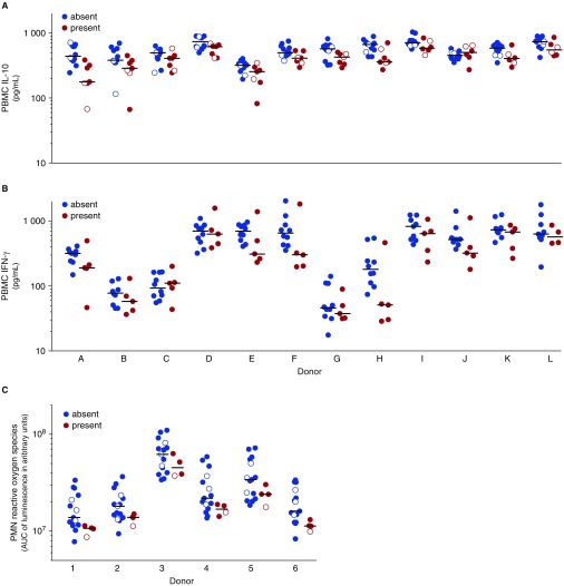
Figure 3.
In vitro responses of selected assays for targets of independent mutation. Stimulation was performed with lysate of Mycobacterium tuberculosis strains from lineage 1 (filled circles) and lineage 4 (open circles) that did not harbor (blue) or harbored (red) a mutation in (A) espE, (B) PE-PGRS56, or (C) Rv2813–2814c. Peripheral blood mononuclear cells (PBMCs) of 12 healthy donors (A–L) were stimulated. (A) IL-10 was measured after 24 hours. (B) IFN-γ was measured after 7 days. (C) Polymorphonuclear cells (PMNs) of six healthy donors (1–6) and reactive oxygen species were measured by luminol-enhanced chemiluminescence and plotted in arbitrary units of the area under the curve (AUC) of the measurement over the first hour after stimulation. Circles in A show overlap because of limited variation; only lineage 1 strain results are shown in B, because no mutations occurred in PE-PGRS56 genes in strains from lineage 4.

