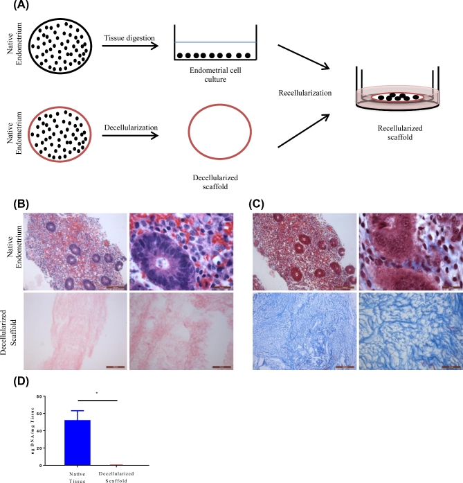
Figure 1.
Diagrammatic representation of the development of the 3D recellularized endometrial culture system. Native endometrial tissue were either digested for cell isolation or prepared for decellularization. Isolated cells were allowed to expand for 5–7 days on 2D plates. Expanded endometrial cells were trypsinized and reseeded on decellularized scaffolds placed inside 12-mm-diameter inserts and cultured in a 24-well plate (A). Representative microscopic images (10× and 100×) of native human endometrium (top row) and decellularized scaffolds (bottom row). Cellular components were stained by H&E (B). ECM of native endometrium and decellularized scaffolds was stained with trichrome staining (C). DNA was isolated from matched native endometrium and decellularized scaffolds and measured n = 3 (D). Data are shown as mean ± standard deviation. *P < 0.05 error bar indicates statistically significant difference between native endometrium and decellularized scaffold. Scale bars represent 100 and 20 μm, respectively.

