
Figure 2.
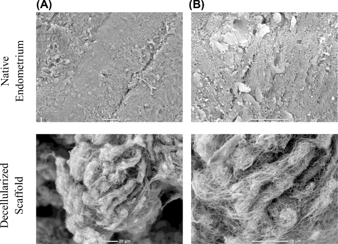
SEM images of normal endometrium and decellularized scaffolds. Native endometrial tissue or decellularized scaffold were fixed in 10% buffered formalin and processed for SEM at (A) 650× (B) 1500×. Scale bars represent 20 μm.
Official websites use .gov
A
.gov website belongs to an official
government organization in the United States.
Secure .gov websites use HTTPS
A lock (
) or https:// means you've safely
connected to the .gov website. Share sensitive
information only on official, secure websites.
SEM images of normal endometrium and decellularized scaffolds. Native endometrial tissue or decellularized scaffold were fixed in 10% buffered formalin and processed for SEM at (A) 650× (B) 1500×. Scale bars represent 20 μm.