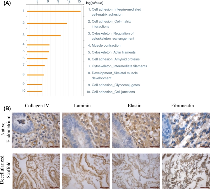
Figure 3.
Proteomics analysis and immunohistochemical staining of retained ECM proteins in decellularized scaffolds. Gene ontology analysis was performed to determine the top processes enriched in the decellularized matrix from three patients (A). IHC staining IV, pan-laminin, elastin and fibronectin in native endometrium (top panel) and decellularized scaffolds (bottom panel) are shown (B). Scale bars represent 20 μm.

