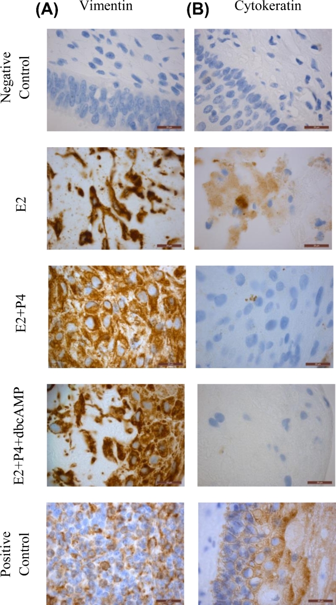
Figure 4.

Recellularization of endometrial scaffold and treatment with a 28-day stepwise hormone protocol. At the end of the experiment, immunohistochemical staining of vimentin (A) and cytokeratin (B) was performed to confirm the presence of stromal and epithelial cells respectively. Representative images from recellularized scaffolds using cells from patients in the proliferative phase are shown (n=3). One recellularized scaffold per patient was used in each treatment group. Scale bars represent 20μm.
