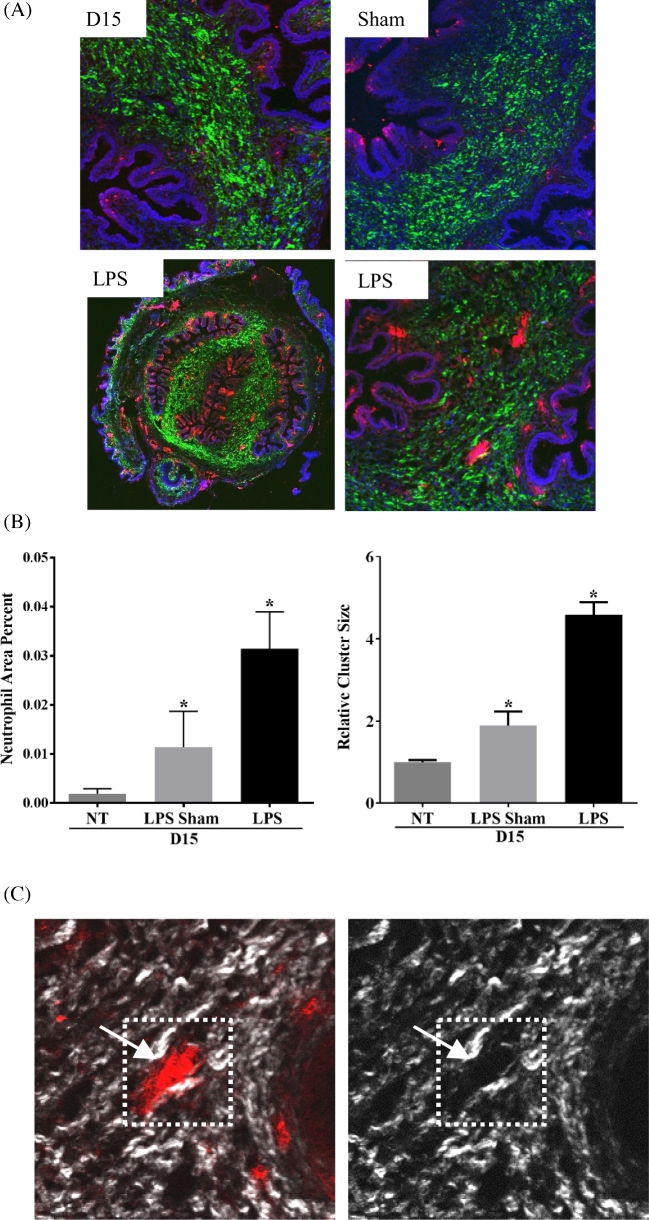
Figure 7.
Cervix from LPS-treated mice contains increased neutrophil clusters concentrated in the SE stromal regions. (A) Dual imaging of collagen by SHG (green), neutrophils (red) by IF using the antibody 7/4. High-magnification images in top panel and lower right to day 15, day 15 sham, and day 15 LPS cervices. Lower left panel is a low-magnification tiled image from day 15 LPS. (B) Quantification of neutrophil cluster size and area percent of neutrophils. Both parameters were significantly increased in LPS-treated cervix when compared to day 15 untreated (NT) and day 15 sham cervix (day 15 n = 3, LPS sham n = 2, and LPS n = 5; *P < 0.05). (C) Dual SHG (white) and IF—7/4 (red) images. The left panel indicates a neutrophil cluster and the right panel indicates reduced SHG signal in the region with neutrophil cluster.

